Karuna Yoga Vidya Peetham Bangalore

karuna yoga vidya peetham logo

Blog

-
Blog

Siddhasana (Adept’s Pose)

Siddhasana (Adept’s Pose) Yonisthanakam angghrimulaghatitam   Krtva drdham vinyaset / Mendhre padam  athaikameva hrdaye Krtva hanum susthiram // Sthanuh samyamitendriyoa chaladrsa pasyed bhruvor antaram/ Hyetan mokshakapatabhedajanakam Siddhasanam prochyate//(Chapter -1, Verse -35) Press the perineum with the heel of one foot, place the other foot on top of the genitals. Having done this, rest the chin on to the chest. Remaining still and steady, with the senses controlled, gaze steadily into the eyebrow center; it breaks open the door to liberation. This is called siddhasana. (Chapter -1, Verse -35)

Read More »

Shavasana (Corpse Pose)

Shavasana (Corpse Pose) Uttanam sabavad bhumau  sayanam tat savasanam/ Savasanam srantiharam chittavisrantikarakam//(Chapter -1, Verse 32). Lying flat on the ground with the face upwards, in the manner of a dead body, is savasana. It removes tiredness and enables the mind-body complex to relax deeply. (Chapter -1, Verse 32).

Read More »

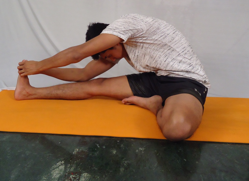
Paschimottanasana (Back Stretching Pose)

Paschimottanasana (Back Stretching Pose) Prasarya padau bhuvi dandarupau Dorbhyam padaghradvitayam ghrhitva/ Januparinyastalalatadeso Vasedidam paschimatanamahuh.// (Chapter -1, Verse 28). Stretching the legs in front on the ground, like a stick; bending forward, holding the toes with both hands and placing the forehead on the knees, is known as Paschimottanasana. (Chapter -1, Verse 28). Paschimottanasana Iti paschimatanamasanaghryam Pavanam paschimavahinam karoti/ Udayam jatharanalasya kuryad Udare karsyamaroghatam cha pumsam//(Chapter -1, Verse 29). Paschimottanasana is the best among asanas. By this asana the pranic currents rise through sushumna, the digestive fire stimulates, the abdomen becomes flat, and the practitioner becomes free from all kind of diseases. (Chapter -1, Verse 29).  

Read More »

Padmasana (lotus pose)

Padmasana (lotus pose) Vamorupari dakshinam cha charanam Samsthapya vamam tatha / Dakshorupari paschimena vidhina Dhrtva karabhyam drdham // Angghushthau hrdaye nidhaya Chibukam nasaghramalokayet / Etadvyadhivinasakari yaminam Padmasanam prochyate//(Chapter -1, Verse 44) Place the right foot on the left thigh and the left foot on the right thigh, cross the hands behind the back and firmly hold the toes. Press the chin against the chest and look at the tip of the nose. This is called padmasana, the destroyer of a yogi’s diseases. (Chapter -1, Verse- 44). Uttanau charanau krtva urusamsthau prayatnatah / Urumadhye tathottanau pani krtva tato drsau//(Chapter -1, Verse-45) Place the feet on the thighs, soles upward, palms in the middle of the groin, facing upward. (Chapter -1, Verse-45)   Nasaghre vinyasedrajadantamule tu jihvaya/ Uttambhya chibukam vakshasyutthapy pavanam sanaih//(Chapter -1, Verse-46) Gaze at the nose tip, keeping the tongue pressed against the root of the upper teeth and the chin against the chest, and slowly raise the prana upward. (Chapter -1, Verse-46) Idam padmasanam proktam sarvavyadhivinasanam/ Durlabham yena kenapi dhimata labhyate bhuvi//(Chapter -1, Verse-47) This is called padmasana, destroyer of all diseases. Ordinary people cannot achieve this posture, only the few wise ones on this earth can do.    (Chapter -1, Verse -47).

Read More »

Mayurasana (Peacock Pose)

Mayurasana (Peacock Pose) Dharam avashtabhya karadvayena Tatkurparasthapitanabhiparsvah/ Uchchasano dandavad utthitah san Mayuram etat pravadanti pitham//(Chapter -1, Verse 30).   Lie down on the stomach, placing both hands on the ground under the body and place the elbows at the sides of the navel. Raise the body high, keeping it like a stick. This is called  peacock pose by the expert Yogis. (Chapter -1, Verse 30).   Harati sakalaroghanasu ghulmodaradin Abhibhavati cha doshanasanam srimayuram/ Bahu kadasanabhuktam bhasma kuryadasesham Janayati // jatharaghnim jarayetkalakutam/(Chapter -1, Verse 31).   Mayurasana quickly alleviates all diseases like enlargement of the glands, dropsy and other stomach disorders. It rectifies imbalance of the humors (vata, pitta, kapha). It reduces to ashes all food taken indiscriminately, kindles the gastric fire and enables destruction of deadly poison). (Chapter -1, Verse 31).  

Read More »

Matsyendrasana (Spinal Twist Pose)

Matsyendrasana (Spinal Twist Pose) Vamoru mularpitadakshapadam Janor bahir veshtitavamapadam/ Praghrhya  tishthet parivartitangghah srimatysanathoditam asanam syat//(Chapter -1, Verse 26).   Place the right foot at the base of the left thigh, the left foot at the side of the right knee. Take hold of the left foot with the right hand, pass the left arm behind the waist and remain with the body turned. This asana is explained by Sri Matsyendranath. (Chapter -1, Verse 26). Matsyendrapitham jatharapradiptim Prachandarughmandalakhandanastram/ Abhyasatah kundaliniprabodham Chandrasthiratvam cha dadati pumsam//(Chapter -1, Verse 27). Practice of this asana Matsyendrasana increases the digestive fire to such an incredible capacity, this will eradicate all stomach diseases and thus awakening the serpent power and bringing equilibrium in the Bindu. (Chapter -1, Verse 27).

Read More »

Kukkutasana (Cockerel Pose)

Kukkutasana (Cockerel Pose) Padmasanam tu samsthapya janurvorantare karau / Nivesya bhumau samsthapya vyomastham kukkutasanam//(Chapter -1, Verse 23).   By adopting Padmasana, insert the hands between the thighs and calves, planting them the hands firmly on the ground; raise the body in the air. This is called as Kukkutasana. (Chapter -1, Verse 23).

Read More »

 Koormasana (Tortoise Pose)

 Koormasana (Tortoise Pose) Ghudam nirudhya ghulphabhyam vyutkramena samahitah/ Kurmasanam bhavedetaditi yogavido viduh//(Chapter -1, Verse 22).   Press the anus firmly with the ankles in the opposite direction and sit well-poised. According to the Yogis this is called as Koormasana. (Chapter -1, Verse 22).  

Read More »

Gomukhasana (Cow’s Face Pose)

Gomukhasana (Cow’s Face Pose) Savye dakshinaghulpham tu prshthaparsve niyojayet / Dakshineapi tatha savyam ghomukham ghomukhakrtih//(Chapter -1, Verse 20).   Place the right ankle next to the left buttock and the left ankle next to the right buttock. This is gomukhasana and it looklike the face of the cow. (Chapter -1, Verse 20).

Read More »

Dhanurasana (Bow Pose)

 Dhanurasana (Bow Pose) Padanghushthau tu panibhyam ghrhitva sravanavadhi / Dhanurakarshanam kuryad dhanurasanam uchyate//(Chapter -1, Verse 25).   Holding the toes with the hands, pull them up to the ears as if drawing a bow. This is called Dhanurasana. (Chapter -1, Verse 25).

Read More »

Bhadrasana (gracious pose)

Bhadrasana (gracious pose) Ghulphau cha vrshanasyadhah Sivantyah parsvayoh kshipet / Savyaghulpham tatha savye Dakshaghulpham tu dakshine // Parsvapadau cha panibhyam Drdham baddhva sunischalam / Bhadrasanam bhavedetatsarvavyadhivinasanam //(Chapter -1, Verse- 53).   Place the ankles below the genitals on the sides by the perineum, left ankle on the left (side) right ankle on the right (side).(Chapter -1, Verse- 53). Ghorakshasanamityahuridam vai siddhayoghinah/ Evamasanabandheshu yogindro vighatasramah // Abhyasennadikasuddhim mudradipavanakriyam/(Chapter -1, Verse-54).   Then hold the feet, which are on their sides, firmly with the hands and remain motionless. This is bhadrasana which destroys all diseases. The yogis who are perfected (siddhas) call it gorakshasana. (Chapter -1, Verse-54).

Read More »

Who can practice Yoga?

Hatha Yoga Pradipika – Who can practice Yoga? Yuvo vrddhoativrddho va vyadhito durbaloapi va / Abhyasat siddhim apnoti sarvayogheshvatandritah//(Chapter -1, Verse -64). Either, Young or old, very old, sick or feeble, one can attain perfection in all the Yogas by regular constant practicing. (Chapter -1, Verse – 64).  

Read More »

Food conducive for Hatha Yogic Practices

Hatha Yoga Pradipika – Food conducive for Hatha Yogic Practices Ghodhuma-sali-yava-shashtika-sobhanannam / Kshirajyakhanda-navanitasi hamadhuni// Sunthipatolakaphaladikapanchasakam / Mudghadidivyam udakam cha yamindrapathyam//(Chapter -1, Verse -62). The most supporting foods for the Hatha Yogic practices are: good grains, wheat, rice, barley, milk, ghee, brown sugar, sugar candy (crystallized sugar), honey, dry ginger, patola fruit (species of cucumber), five vegetables, mung, pulses, and pure water. (Chapter -1, Verse -62). Pushtam sumadhuram snighdham Gavyam dhatupraposhanam / Manobhilashitam yoghyam yogi bhojanamacharet//(Chapter -1, Verse-63). The Hatha Yogi should take nourishing and sweet food mixed with, ghee and milk; it should nourish the dhatus (basic body constituents) and be pleasing and suitable for body-mind-soul. (Chapter -1,Verse-63).  

Read More »

Food Prohibited for Hatha Yogic Practices

Hatha Yoga Pradipika – Food Prohibited for Hatha Yogic Practices Katvamla-tikshna-lavanoshna-haritasaka / Sauvira-taila-tila-sarshapa-madya-matsyan // Ajadi-mamsa-dadhi-takra-kulattha-kola / Pinyaka-hingghu-lasunady-amapathyam-ahuh//(Chapter -1, Verse- 59). The foods which prohibited for the Hatha Yogic practitioners are: those which are bitter, sour, pungent, salty, heating, green vegetables, sour gruel, oil, sesame and mustard, alcohol, fish, flesh foods, curds, buttermilk, horse gram, fruit of jujube, oil cakes, asafetida and garlic. (Chapter -1, Verse- 59). Bhojanam ahitam vidyāt punarasyoshnīkrtam rūksham/ Atilavanamam layuktam kadaśanaśākotkam varjyam// (Chapter -1, Verse -60).   Unnourishing food should not be consumed, that which is reheated after becoming cold, which is dry (devoid of natural oil), which is excessively salty or acidic, stale or has too many mixed vegetables. (Chapter -1, Verse -60).  

Read More »

Moderate diet for Hatha Yogic Practices

Hatha Yoga Pradipika -Moderate diet for Hatha Yogic Practices Susnighdhamadhuraharaschaturthamsavivarjitah / Bhujyate sivasamprityai mitaharah sa uchyate//(Chapter -1, Verse -58). Mitahara is defined as agreeable and sweet food, leaving one fourth of the stomach free, and eaten. (Chapter -1, Verse-58).

Read More »

Electrocardiogram ECG

Electrocardiogram ECG: The ECG shows the heart’s electrical activity as line tracings on paper. It’s the recording of electrical activity of the heart. ECG is an instrument which is used to record the electrical current generated in the heart. By means of this instrument, the electrical current generated in the heart is conducted to remote by connecting any two parts of the body with this instrument. The connections are called as leads they are. A normal ECG contains waves, intervals, segments and one complex, as defined below. Wave: A positive or negative deflection from the baseline that indicates a specific electrical event. The waves on an ECG include the P wave, Q wave, R wave, S wave, T wave and U wave. Interval: The time between two specific ECG events. The intervals commonly measured on an ECG include the PR interval, QRS interval (also called QRS duration), QT interval and RR interval. Segment: The length between two specific points on an ECG that are supposed to be at the baseline amplitude (not negative or positive). The segments on an ECG include the PR segment, ST segment, and TP segment. Complex: The combination of multiple waves grouped together. The only main complex on an ECG is the QRS complex. The P wave indicates atrial depolarization. The QRS complex consists of a Q wave, R wave, and S wave and represents ventricular depolarization. The T wave comes after the QRS complex and indicates ventricular repolarization.  

Read More »

Functions of the Skeletal System

Functions of the Skeletal System? Support of the body Locomotion Provide protection for internal organs Act as a site for the production of blood cells, Calcium storage   & endocrine regulation. Growth and development  

Read More »

Disorders of blood vessels

Disorders of blood vessels: Arteriosclerosis: Arteriosclerosis is the thickening, hardening, and loss of elasticity of the walls of arteries. This process gradually restricts the blood flow to one’s organs and tissues. Atherosclerosis: it’s a thickening of arterial walls due to the deposition of fat and white blood cells). An aneurysm: it’s a local abnormal dilation of an artery. Classified into 3 types: fusiform, saccular, and dissecting. Embolus: it’s a clot moving in circulation and becomes impacted in a small vessel. Thrombus: it’s a clot obstructing a blood vessel at the point where it’s actually formed. Venous thrombosis: its blood clot obstructing a vein. Thrombophlebitis: it’s inflammation of vein complicated by an obstructing clot. Varicose vein: it’s a dilated and tortuous superficial vein. This occurs because valves in the veins don’t close. So, the backward flow of blood in the vein is not prevented. This leads to dropping back of the column occurs in 1- long saphenous vein. 2- short saphenous vein. 3- anterior tibial vein.  

Read More »

Properties of skeletal muscle

Properties of skeletal muscle: Excitability and irritability: It’s the property of a muscle to respond to a stimulus. If the response occurs in the front of a contraction. It’s called as contractility. Tonicity: Muscles of a living organism are in the partially contracted state, even at rest. They are never fully relaxed and they will resist being stretched the resistance to stretch is called as tonicity. All or none response: When a stimulus is given to a muscle, the muscle either contracts to the maximum or it doesn’t contract at all. When a weak stimulus is applied to a muscle, it doesn’t produce any response. But when the stimulus is gradually increased, the muscle contracts at a point to the maximum level. Later even a greater degree of stimulus does not increase the degree of contraction. Summation: When a second stimulus is given to a muscle even while it’s contracting due to the first stimulus, the degree of contraction is more. This phenomenon is called as a summation. The increased response is due to stimulation of more and more motor units by repetitive stimuli. Treppe or staircase phenomenon: Stimulation of a muscle at regular short intervals increases the amplitudes of contraction *which goes on increasing like the steps of a staircase*. This occurs due to increased irritability of the muscle produced by chemical products of the earlier contractions. Tetanus: When a muscle is stimulated repeatedly at a very faster rate, the muscle contracts maximally. The muscle remains at this maximal state of contraction till 1- the stimulus continues or 2- it gets fatigued. This sustained maximal contraction is called tetanus. Refractory period: Muscle, like other excitable tissues, has a period following its action potential during which the membrane will not respond to stimulation regardless of the strength. Isotonic contraction: It’s the contraction in which the muscle shortens under constant load. It occurs in muscles during walking, running or lifting. Isometric contraction: In this type of contraction, the muscle develops tension but it does not shorten in length e.g. maintaining the posture against gravity. Fatigue: It’s a state of reduced excitability and contractility of a muscle. It’s produced by rapid and repeated stimulation of the muscle. Fatigue may occur due to depletion of energy and accumulation of metabolites like lactic acid.  

Read More »

Joint Disorders

Joint Disorders Arthritis – inflammation of a joint. Characterized by pain, stiffness, and swelling. Over time, the joint can become severely damaged. Osteoarthritis is the most common type of arthritis. It’s often related to aging or to an injury. Rheumatoid arthritis is the most common form of this kind of Autoimmune arthritis. Rheumatoid arthritis happens when your body’s immune system attacks healthy cells in your body by mistake. Bursitis – inflammation of a fluid-filled sac that cushions the joint, Excessive use of a joint or stress on a bursa may cause bursitis, an inflammation of a bursa. Joint Dislocation: Luxation or Dislocation, displaces the articulating bones of a joint and usually results from a fall or other unusual body movement. Joint dislocation produces an obvious deformity of the joint, some loss of ability to move the articulated bones, localized pain, and swelling. Sprains: Sprains result from overstretching or tearing the connective tissues, ligaments, and tendons associated with a joint, but they do not dislocate the articular bones.  

Read More »

Cardiac Cycle

The Cardiac cycle:   the function of the heart is to maintain a constant circulation of blood throughout the body. It acts as a pump to maintains a constant circulation of blood throughout the body. It’s achieved as follows: The functions of the cardiovascular system are to provide the tissues of the body with oxygen and nutrients which are transported in the blood. The superior and inferior vena cava veins bring venous blood from various body parts to the heart. This venous blood fills the right atrium. When it’s full, the right atrium contracts sending blood to the right ventricle. Now the right ventricle contracts this sends blood to the lungs through pulmonary trunk which divides into right and left pulmonary arteries.  The blood gets oxygenated in the lungs and then the oxygenated blood is carried by a pulmonary vein to the left atrium. Now the left atrium contracts and sends blood to the left ventricle. The left ventricle contracts and send blood into the aorta, this blood circulated throughout the body. This is achieved by rhythmic contraction of its muscle. The cardiac cycle is the sequence of events which occur in the heart during a single beat. Heart rate is between 60 to 90 normally. With an average rate of 72. So the time taken for one beat is 0.8 second. So the sequence of events occurs every 0.8 seconds. The cardiac cycle occurs in 2 phases: Systole which means a period of contraction. Diastole which means a period of relaxation. The cycle of events occurs as follows: To start with blood from the veins fill the 2 atria, the superior and inferior vena cava fill the right atrium, the pulmonary veins fill the left atrium. This is followed by a wave of contraction in the atria, this leads to emptying of atrial blood into the respective ventricle. When the ventricles are full, they contract, now the blood present in ventricles is forced into systemic and pulmonary circulation. At this stage, the semilunar valves guarding the aorta and pulmonary arteries are opened. At the same time, the atrioventricular are opened. At the same time, the atrioventricular valves are closed. This period of muscular contraction of the heart is called systole. This is followed by a period of relaxation called diastole during which there’s no contraction. This sequence of events constitutes the cardiac cycle.  

Read More »

Somatic Nervous System

Somatic Nervous System The somatic system is the part of the peripheral nervous system that is responsible for carrying motor and sensory information both to and from the central nervous system. This system is made up of nerves that connect to the skin, sensory organs, and all skeletal muscles. The system is responsible for nearly all voluntary muscle movements as well as for processing sensory information that arrives via external stimuli including hearing, touch, and sight. The somatic system is responsible for transmitting sensory information as well as for vol untary movement. This system contains two major types of neurons: Sensory neurons (or afferent neurons) that carry information from the nerves to the central nervous system. It is these sensory neurons that allow us to take in sensory information and send it to the brain and spinal cord. Motor neurons (or efferent neurons) that carry information from the brain and spinal cord to muscle fibers throughout the body. These motor neurons allow us to take physical action in response to stimuli in the environment.

Read More »

Skeletal System

Skeletal system  The human skeleton is made up of 206 bones making the human body a multifunctional structure. The skeletal system is composed of Bones, Joints, Cartilage, Tendons, and Ligaments. Human infants are born with about 270 bones, most of which ossifies as they grow up. The skeletons of adult males and females have some variation, primarily to accommodate childbirth. The female pelvis is flatter, more rounded and proportionally larger. Bone: A rigid form of connective tissue that is part of the skeletal system of vertebrates and is composed principally of calcium phosphate and calcium carbonate. It also serves as a storage area for calcium, playing a large role in calcium balance in the blood. The ligament is a small band of dense, white, fibrous elastic tissue. Ligaments connect the ends of bones together in order to form a joint. They also assist in holding organs in place. Tendons are a tough, flexible and inelastic band of fibrous connective tissue that connects muscles to bones and is capable of withstanding tension. Tendons are similar to ligaments; both are made of collagen.  

Read More »

Respiratory System

Respiratory system The Human Respiratory System is a series of organs which facilitates the diffusion of oxygen into the bloodstream and distributes throughout the body and receives carbon dioxide from the blood and removes the process of exhalation. The primary organs of the respiratory system are lungs, which carry out this exchange of gases as we breathe. The respiratory system consists of the upper and lower respiratory tracts. The upper respiratory tract composes of Nasal cavity, Pharynx, and Larynx. The lower Respiratory Tract composes of Lungs, Diaphragm, Trachea, Bronchi and Bronchioles. Gaseous Exchange: The main function of the respiratory system is a gaseous exchange. This refers to the process of Oxygen and Carbon Dioxide moving between the lungs and blood. Diffusion occurs when molecules move from an area of high concentration (of that molecule) to an area of low concentration. This occurs during the gaseous exchange as the blood in the capillaries surrounding the alveoli has a lower oxygen concentration of Oxygen than the air in the alveoli which has just been inhaled.  

Read More »

Physiology of Muscle contraction

Physiology of Muscle contraction: Muscles contract to produce force, the actin and myosin filaments within the sarcomeres of muscle fibers bind to create cross-bridges and slide past one another, creating a contraction. The events of muscle contraction: During a normal resting state of a muscle, the muscle membrane is in a polarized state, this occurs because: The interior of the muscle cell is negatively charged. It contains a large concentration of potassium ions. The exterior of the muscle cell is positively charged. It contains a large contraction of sodium ions. For a contraction to occur there must first be a stimulation of the muscle in the form of an impulse from a motor neuron. A motor neuron stimulates a number of muscle fibers within a muscle. The Nerve impulse reaches the muscle fibers of a Neuromuscular Junction, it stimulates a reaction in each sarcomere between the actin and myosin filaments.  This reaction results in muscular contraction. When the muscle contraction is over, acetylcholine is destroyed by the acetyl cholinesterase. Now, Potassium ions move into the cell and sodium ions get out of the cell. ATP is re-synthesized allowing actin and myosin to maintain their strong binding state. This produces repolarization of the muscle membrane. Now the muscle is ready to contract again.  

Read More »

Disorders of blood pressure

Disorders of blood pressure Hypertension’s Hypertension is a rise in blood pressure above normal. It’s difficult to define the average normal blood pressure. It varies from one age group to other. The blood pressure in an adult may be 120/80. Hypertension has a strong relation with life style regardless of gender and socio-economic status. The systolic blood pressure(BP) greater than or equal to140 mmHg and/or diastolic BP greater than or equal to 90 mmHg is currently the widely accepted diagnostic criteria for hypertension. The management of hypertension is a major public health challenge in developing countries, even though a wide range of antihypertensives are developed and prescribed for the management of hypertension. Hypertension is a major risk factor for stroke, myocardial infarction, vascular diseases and kidney disorders. Hypotension: Hypotension is low blood pressure which can be physiological in health during rest and in elderly. Mostly it occurs as a complication of other diseases e.g. shock, myocardial infarction, and hemorrhage. Blood pressure varies from one person to another. A drop as little as 20 mmHg, can cause problems for some people. Low blood pressure leads to inadequate blood supply to the brain. It may produce brief unconsciousness, fainting. if this is prolonged, death may occur. Severe hypotension can be caused by sudden loss of blood (shock), severe infection, heart attack, or severe allergic reaction.  

Read More »

Neuromuscular junction (Myoneural junction)

Neuromuscular junction (Myoneural junction) A neuromuscular junction is a synapse between a motor neuron and skeletal muscle. The space between the motor neuron and the skeletal muscle cell is simply referred to as a synapse. Synaptic transmission includes all the events within the synapse leading to excitation of the muscle. At this site, the nerve loses its myelin sheath and gets expanded. Acetylcholine is stored in some vesicles present in this expanded portion. Motor end plate is the part of the muscle facing the expanded nerve terminal. The neuron is sending the transmission and is thus referred to as the pre-synaptic cell, while the muscle is receiving the transmission and is referred to as the post-synaptic cell. Neurotransmitters are molecules stored in the pre-synaptic cell that is secreted into the synapse. Neurotransmitters, in turn, bind to receptors on the postsynaptic cell membrane, and these receptors are specific for that neurotransmitter. The structure motor end plates convoluted and it has enzyme acetyl cholinesterase.  

Read More »

Common Bone Disorders

Common Bone Disorders Osteoporosis- Osteoporosis is a common disease that weakens bones, characterized with the prominent porous formation in the shaft or articulating part of the bone. As bones weaken, your risk of sudden and unexpected fractures increases. Rickets- Rickets is a skeletal disorder that results from a lack of vitamin D, calcium, or phosphate. Osteomyelitis- Osteomyelitis is a bone infection that is caused by bacteria, characterized by severe pain and tenderness. Osteosarcoma- An osteosarcoma or osteogenic sarcoma is a cancerous tumor in a bone. Specifically, it is an aggressive malignant neoplasm that arises from primitive transformed cells of mesenchymal origin and that exhibits osteoblastic differentiation and produces malignant osteoid. Osteomalacia- Osteomalacia is softening of the bones. It most often occurs because of a problem with vitamin D.

Read More »

Respiration Mechanism

Respiration mechanism Normal breathing involves several different mechanisms. Shallow breathing is accomplished by the contraction of the diaphragm and the external intercostal muscles for inhalation. During exhalation, the muscles relaxing and the elasticity of the lungs returning to their resting volume expels air out of the lungs.  Deep Breathing is accomplished by an inferior movement of the diaphragm towards the abdomen. The external intercostal muscles along with the sternocleidomastoid and scalene muscles in the neck expand the space between the ribs, increasing the volume of the chest. During deep exhalation, the internal intercostal muscles and abdominal muscles contract to decrease the volume of the thoracic cavity, forcing air out of the lungs. Breathing is controlled by the brain and may be controlled both consciously and unconsciously. Unconscious control of breathing is maintained by the respiratory center of the brainstem, which monitors the concentration of gases in the blood and adjusts the rate and depth of breathing as needed. During Yoga practice, the respiratory center automatically increases the breathing rate to provide constant levels of oxygen to the blood. During rest, the respiratory center reduces the breathing rate to prevent hyperventilation and maintain healthy oxygen and carbon dioxide levels in the blood. Conscious control of breathing is maintained by the cerebral cortex of the brain. The cerebral cortex may override the respiratory center during the practice of Pranayama. Unconscious control of breathing resumes as soon as conscious control of breathing ends, preventing the body from suffocating from lack of breathing.  

Read More »

Peripheral Nervous System

Peripheral Nervous System: The Peripheral nervous system (PNS) is the division of the nervous system containing all the nerves that lie outside of the central nervous system (CNS). The primary role of the PNS is to connect the CNS to the organs, limbs, and skin. These nerves extend from the central nervous  system to the outermost areas of the body. The peripheral system allows the brain and spinal cord to receive and send information to other areas of the body, which allows us to react to stimuli in our environment. The nerves that make up the peripheral nervous system are actually the axons or bundles of axons from neuron cells. In some cases, these nerves are very small but some nerve bundles are so large that they can be easily seen by the human eye.  

Read More »

Skeletal Muscle Structure

Skeletal Muscle Structure Skeletal muscles are composed of a large number of muscle fibers. Each muscle fiber has no one or more nuclei which lie in the periphery. The cytoplasm of the muscle cell is as sarcoplasm. Skeletal muscles are composed primarily of contractile material. Skeletal muscle is a composite tissue of connective tissue, blood vessels, and nerves as well as contractile material, these “minor” tissues may strongly influence muscle function. Myofibrils are very small parallel filaments which lie in the cytoplasm. The membrane of the muscle fiber is called as sarcolemma. Each muscle fiber is embedded in a connective tissue called endomysium. The fibers within a muscle are arranged in bundles. Each bundle is enclosed in a sheath called perimysium.  

Read More »

Conducting system of the heart

Conducting system of the heart Conducting system of the heart: the impulse for cardiac contraction is transmitted through the conduction system of the heart. This system is made of : Sino atrial node (SA-node) Atrioventricular node (AV-node) Bundle of his Purkinje fibers. Heart sounds:  Totally 4 sounds are produced by the heart. The first sound as LUB and the second sound as DUB can be heard with a stethoscope. The third and fourth sounds cannot be heard. The “ lub” is the first heart sound, commonly termed S1, and is caused by turbulence caused by the closure of mitral and tricuspid valves at the start of systole. The second sound, “dub” or S2, is caused by the closure of aortic and pulmonic valves, marking the end of systole. Heart Murmurs. The most common abnormal heart sound is a heart murmur. A murmur is a blowing, whooshing, or rasping sound that occurs during your heartbeat. Pulse: its throbbing sensation felt over the walls of arteries. It’s defined as the pressure difference transmitted in the form of a wave over the arterial walls. Heart Rate: the normal resting heart rate for adults ranges from 60 to 100 beats a minute. The wrist is the common site where the pulse is usually felt. At this site, the radial artery is very superficial. This pulse is called as a radial pulse. Other arteries where a pulse can be felt are a carotid artery. Facial artery and temporal artery. In case of fever, the rate of pulse increases at the rate of 10 per every rise of 1F.Generally, veins don’t exhibit pulsation. The only vein which exhibits pulsation is jugular vein which is nearer to the heart.  

Read More »

Classification of Human Bones

Classification of Bones according to shape Long bones: Long bones are characterized by a long tubular shaft and an articular surface at each end of the bone where ligaments and tendons attach. They’re found in the limbs to facilitate movement and support the weight of the body. These bones include the major bones of the arms and legs such as the humerus and femur, tibia and fibula, and the radius and ulna. Short bones: Short bones are roughly cube shaped and are as long as they are wide. It has no shaft but it contains a spongy substance covered by a shell of the compact. Helps to provide stability and movement within the ankle and wrist joints. They provide little to no movement. Examples of this type of bone include the carpals and metacarpals in the wrists and ankles. Flat bones: The primary purpose of this type of bone is to protect internal organs such as the brain, heart, and lungs. It also provides a large surface area for muscles to attach to. Examples of this type of bone include the cranium (skull), the thoracic cage (sternum and ribs) and the ilium (pelvis).  These contain two layers of compact bone with a spongy substance in between. Irregular bones: This type doesn’t fall in any category e.g. vertebrae and the bones of the face. These bones vary in size and structure with the shape usually being very complex. Irregular bones serve different functions depending on location. Sesamoid bones: they are small bones which develop in the tendons of muscles e.g. patella of the knee joint. The function of this bone is to protect tendons and diminish friction and wear on joint surfaces.  

Read More »

Pulmonary Ventilation

Pulmonary Ventilation Smooth muscle cells in the walls of the bronchioles adjust their diameter and help to control the flow of air into the alveoli of the lungs. The muscles of the thoracic cavity, including the diaphragm and intercostal muscles, change the volume of the thoracic cavity to force air into and out of the lungs. The diaphragm contracts and pushes itself into the abdominal cavity, increasing the volume of the thoracic cavity and expanding the lungs to draw in fresh atmospheric air. When it relaxes, the diaphragm returns to its original position and forces air out of the lungs during exhalation. The intercostal muscles also aid in respiration by lifting the ribs during deep inhalation to increase the size of the thoracic cavity and by pushing the ribs together during deep exhalation to decrease the thoracic cavity.  

Read More »

Nervous System

Nervous System The nervous system has been divided into two components: The central nervous system which is composed of the brain and the spinal cord, and the peripheral nervous system, which is composed of ganglia and peripheral nerves that lie outside the brain and spinal cord. The peripheral nervous system has been, in turn, divided into two subsystems: Somatic and autonomic. Somatic Nervous system (SNS): The somatic nervous system is responsible for the movement of voluntary muscles and the process known as a reflex arc. This system carries nerve impulses back and forth between the central nervous system and the skeletal muscle’s, skin and the sense organs. Autonomic Nervous system (ANS): The Autonomic Nervous system controls and regulates the internal organs without any conscious recognition or effort by the organism.  

Read More »

CHARACTERISTICS OF SKELETAL MUSCLES

CHARACTERISTICS OF SKELETAL MUSCLES The four major functional characteristics of skeletal muscle are: Contractility    – The ability to shorten which causes movement of the structures to which the muscles are attached. Excitability      – The ability to respond or contract in response to chemical and/or electrical signals. Extensibility     –  The capacity to stretch to the normal resting length after contracting Elasticity          – The ability to return to the original resting length after a muscle has been stretched. Functions of Muscles: Movement: Skeletal muscles move bones by pulling on them and work together to produce smooth movements of our limbs. Mobility is your muscular system’s simplest and most crucial function. Your skeletal muscles are largely responsible for the movements and motions you make. Skeletal muscles are attached to your bones. Maintaining posture, muscle tone, and Stabilizing joints: Skeletal muscles also control posture, from your head down to your toes. Flexibility and strength are keys to maintaining proper posture. Stiff neck muscles, weak back muscles, and tight hip muscles, among other ailments, can throw off your alignment. Producing Heat & Temperature Regulation: most of the heat required for this is produced by muscles, which generate heat as they contract. Skeletal muscle accounts for at least 40% of body mass and is the muscle type responsible for generating most body heat. Circulation: The involuntary cardiac and smooth muscles help your heart beat and blood flow through your body. The cardiac muscle, known as the myocardium, is found in the walls of the heart. The cardiac muscle is controlled by the autonomic nervous system, which is responsible for most bodily functions.  

Read More »

Cardiac Output

Cardiac Output The cardiac output is simply the amount of blood pumped by the heart per minute. Necessarily, the cardiac output is the product of the heart rate, which is the number of beats per minute, and the stroke volume, which is amount pumped per beat. Stroke volume is the amount of blood ejected per beat of the heart. It’s about 70ml. so every mixture about 5040 ml nearly 5 liters of blood is pumped by the heart. Cardiac output depends on the following factors:  

Read More »

Bone structure and formation

Bone structure and formation: Bone is the hardest of the connective tissues. Compact bone (Cortical Bone) and Spongy bone (Spongy Bone) are the two types of osseous tissue or bone tissue that make up bones. The bones are mostly composed of an outer layer of compact bone and an inner layer of spongy bone. Ossification is the process of bone formation. Parts of the skeleton form during the first few weeks after conception. By the end of the eighth week after conception, the skeletal pattern is formed in cartilage and connective tissue membranes and ossification begins.   Compact bone (Cortical Bone): Due to the strength of compact bone, its main functions is to support the entire body. Compact pact bone also stores calcium. While calcium is being stored in the bone it is also the principle molecule that hardens compact bone. Cancellous bone (Spongy Bone): It looks spongy and found in the ends of the long bones, short bones and in between two layers of compact tissue of flat bones. Due to the sponge-like or porous nature of spongy or cancellous bone, spongy bone has a greater surface area compared to compact bone.  This allows the bone marrow to develop in the region of the spongy bone. The branching nature of spongy bone also makes it a prime target in diseases such as osteoporosis.  

Read More »

Lungs Volume

Lungs Volume The total air volume of the lungs is about 4 to 6 liters and varies with a person’s size, age, gender, and respiratory health.   Normal shallow breathing only moves a small fraction of the lungs’ total volume into and out of the body with each breath. This volume of air, known as tidal volume, usually measures only around 0.5 liters. Deep breathing can force more air into and out of the lungs than during shallow breathing. The volume of air exchanged during deep breathing is known as vital capacity and ranges between 3 to 5 liters, depending on the lung capacity of the individual. There is a residual volume of around 1 liter of air that remains in the lungs at all times, even during a deep exhalation.  

Read More »

Central Nervous System

The Central Nervous System: The Central Nervous System comprises of the Brain and the Spinal cord: The Brain plays a central role in the control of most bodily functions, which includes awareness, movements, sensations, thoughts, speech, and memory. The brain takes in sensory information, organizes and synthesizes this input, then provides instructions for motor output to the rest of the body. The brain is the main data center of the body, consisting of the cerebrum (which regulates Cognitive functions) and the cerebellum (which regulates coordination and muscular skeletal functions). The Spinal cord consists of nerves that carry incoming and outgoing messages between the brain and the rest of the body. Spinal cord acts as the center for reflexes. Brain: The human brain is the command center for the human nervous system. It receives input from the sensory organs and sends output to the muscles. The Human Brain weighs about 1.5 kilograms, it makes up about 2 percent of a human’s body weight. The cerebrum makes up 85 percent of the brain’s weight composing of nearly 86 billion neurons. The neurons are the structural and functional unit of the nervous system. The brain develops from 3 sections, they are as follows. Forebrain: also known as prosencephalon, is the anterior part of the brain, includes the cerebral hemispheres, the thalamus forebrain develops into the cerebrum and underlying structures. Midbrain: also known as mesencephalon becomes part of the brainstem, serves important functions like motor activity and sensory functions. Hindbrain: the lower part of the brain stem, comprising the cerebellum, pons cerebelli, and medulla oblongata. Parts of the Brain and functions: AMYGDALA: Lying deep in the center of the limbic emotional brain, this powerful structure, the size, and shape of an almond, is constantly alert to the needs of basic survival. BRAIN STEM: The part of the brain that connects to the spinal cord. The brain stem controls function basic to the survival of all animals, such as heart rate, breathing, digesting foods, and sleeping. It also plays a role in learning. CEREBRUM: This is the largest brain structure in humans and accounts for about two-thirds of the brain’s mass. The two hemispheres of the cerebrum are connected by long neuron branches called the corpus callosum. The cerebrum control most of our body functions such as the state of consciousness, the senses, the body’s motor skills, reasoning, and language. CEREBELLUM: Two peach-size mounds of folded tissue located at the top of the brain stem. It controls most of the learning pathways and coordinated skilled movements. The Frontal Lobe is the most recently-evolved part of the brain. The frontal lobe is dorsolateral prefrontal circuit is the brain’s top executive. It organizes responses to complex problems, plans steps to an objective, searches memory for relevant experience, adapts strategies to accommodate new data, guides behavior with verbal skills and houses working memory. The Temporal Lobe controls memory storage area, emotion, hearing, and, on the left side, language. The Parietal Lobe receives and processes sensory information from the body including calculating location and speed of objects. The Occipital Lobe processes visual data and routes it to other parts of the brain for identification and storage. HIPPOCAMPUS: located deep within the brain, it processes new memories for long-term storage. If you didn’t have it, you couldn’t live in the present, you’d be stuck in the past of old memories. It is among the first functions to falter in Alzheimer’s. HYPOTHALAMUS: Located at the base of the brain where signals from the brain and the body’s hormonal system interact, the hypothalamus maintains the body’s status quo. It monitors numerous bodily functions such as blood pressure and body temperature, as well as controlling body weight and appetite. THALAMUS: Located at the top of the brain stem, the thalamus acts as a two-way relay station, sorting, processing, and directing signals from the spinal cord and midbrain structures up to the cerebrum, and, conversely, from the cerebrum down the spinal cord to the nervous system. Spinal Cord: The spinal cord is a long, fragile tube-like structure that begins at the spinal cord extends from the foramen magnum where it is continuous with the medulla to the level of the first or second lumbar vertebrae. Spinal Cord is a vital link between the brain and the body, and from the body to the brain, which is 40 to 50 cm long and 1 cm to 1.5 cm in diameter. Two consecutive rows of nerve roots emerge on each of its sides, these nerve roots join distally to form 31 pairs of spinal nerves. The spinal cord is a cylindrical structure of nervous tissue composed of white and gray matter, is uniformly organized and is divided into four regions: cervical (C), thoracic (T), lumbar (L) and sacral (S). The spinal cord consists of nerves that carry incoming and outgoing messages between the brain and the rest of the body. It is also the center for reflexes, such as the knee jerk reflex. The spinal nerve roots are formed by the union of dorsal and ventral roots within the intervertebral foramen, resulting in a mixed nerve joined together and forming the spinal nerve. The arterial blood supply to the spinal cord in the upper cervical regions is derived from two branches of the vertebral arteries, the anterior spinal artery, and the posterior spinal arteries. At the level of the medulla, the paired anterior spinal arteries join to form a single artery that lies in the anterior median fissure of the spinal cord. The posterior spinal arteries are paired and form an anastomotic chain over the posterior aspect of the spinal cord. A plexus of small arteries, the arterial vasocorona, on the surface of the cord constitutes an anastomotic connection between the anterior and posterior spinal arteries. This arrangement provides uninterrupted blood supplies along the entire length of the spinal cord. Functions: conducts sensory information from the peripheral nervous system (both somatic and autonomic) to the brain, the spinal nerves carry sensory information (sensations) from the

Read More »
[gravityform id="2" title=false]
Call Back Form