Central Nervous System

The Central Nervous System: The Central Nervous System comprises of the Brain and the Spinal cord: The Brain plays a central role in the control of most bodily functions, which includes awareness, movements, sensations, thoughts, speech, and memory. The brain takes in sensory information, organizes and synthesizes this input, then provides instructions for motor output to the rest of the body. The brain is the main data center of the body, consisting of the cerebrum (which regulates Cognitive functions) and the cerebellum (which regulates coordination and muscular skeletal functions). The Spinal cord consists of nerves that carry incoming and outgoing messages between the brain and the rest of the body. Spinal cord acts as the center for reflexes. Brain: The human brain is the command center for the human nervous system. It receives input from the sensory organs and sends output to the muscles. The Human Brain weighs about 1.5 kilograms, it makes up about 2 percent of a human’s body weight. The cerebrum makes up 85 percent of the brain’s weight composing of nearly 86 billion neurons. The neurons are the structural and functional unit of the nervous system. The brain develops from 3 sections, they are as follows. Forebrain: also known as prosencephalon, is the anterior part of the brain, includes the cerebral hemispheres, the thalamus forebrain develops into the cerebrum and underlying structures. Midbrain: also known as mesencephalon becomes part of the brainstem, serves important functions like motor activity and sensory functions. Hindbrain: the lower part of the brain stem, comprising the cerebellum, pons cerebelli, and medulla oblongata. Parts of the Brain and functions: AMYGDALA: Lying deep in the center of the limbic emotional brain, this powerful structure, the size, and shape of an almond, is constantly alert to the needs of basic survival. BRAIN STEM: The part of the brain that connects to the spinal cord. The brain stem controls function basic to the survival of all animals, such as heart rate, breathing, digesting foods, and sleeping. It also plays a role in learning. CEREBRUM: This is the largest brain structure in humans and accounts for about two-thirds of the brain’s mass. The two hemispheres of the cerebrum are connected by long neuron branches called the corpus callosum. The cerebrum control most of our body functions such as the state of consciousness, the senses, the body’s motor skills, reasoning, and language. CEREBELLUM: Two peach-size mounds of folded tissue located at the top of the brain stem. It controls most of the learning pathways and coordinated skilled movements. The Frontal Lobe is the most recently-evolved part of the brain. The frontal lobe is dorsolateral prefrontal circuit is the brain’s top executive. It organizes responses to complex problems, plans steps to an objective, searches memory for relevant experience, adapts strategies to accommodate new data, guides behavior with verbal skills and houses working memory. The Temporal Lobe controls memory storage area, emotion, hearing, and, on the left side, language. The Parietal Lobe receives and processes sensory information from the body including calculating location and speed of objects. The Occipital Lobe processes visual data and routes it to other parts of the brain for identification and storage. HIPPOCAMPUS: located deep within the brain, it processes new memories for long-term storage. If you didn’t have it, you couldn’t live in the present, you’d be stuck in the past of old memories. It is among the first functions to falter in Alzheimer’s. HYPOTHALAMUS: Located at the base of the brain where signals from the brain and the body’s hormonal system interact, the hypothalamus maintains the body’s status quo. It monitors numerous bodily functions such as blood pressure and body temperature, as well as controlling body weight and appetite. THALAMUS: Located at the top of the brain stem, the thalamus acts as a two-way relay station, sorting, processing, and directing signals from the spinal cord and midbrain structures up to the cerebrum, and, conversely, from the cerebrum down the spinal cord to the nervous system. Spinal Cord: The spinal cord is a long, fragile tube-like structure that begins at the spinal cord extends from the foramen magnum where it is continuous with the medulla to the level of the first or second lumbar vertebrae. Spinal Cord is a vital link between the brain and the body, and from the body to the brain, which is 40 to 50 cm long and 1 cm to 1.5 cm in diameter. Two consecutive rows of nerve roots emerge on each of its sides, these nerve roots join distally to form 31 pairs of spinal nerves. The spinal cord is a cylindrical structure of nervous tissue composed of white and gray matter, is uniformly organized and is divided into four regions: cervical (C), thoracic (T), lumbar (L) and sacral (S). The spinal cord consists of nerves that carry incoming and outgoing messages between the brain and the rest of the body. It is also the center for reflexes, such as the knee jerk reflex. The spinal nerve roots are formed by the union of dorsal and ventral roots within the intervertebral foramen, resulting in a mixed nerve joined together and forming the spinal nerve. The arterial blood supply to the spinal cord in the upper cervical regions is derived from two branches of the vertebral arteries, the anterior spinal artery, and the posterior spinal arteries. At the level of the medulla, the paired anterior spinal arteries join to form a single artery that lies in the anterior median fissure of the spinal cord. The posterior spinal arteries are paired and form an anastomotic chain over the posterior aspect of the spinal cord. A plexus of small arteries, the arterial vasocorona, on the surface of the cord constitutes an anastomotic connection between the anterior and posterior spinal arteries. This arrangement provides uninterrupted blood supplies along the entire length of the spinal cord. Functions: conducts sensory information from the peripheral nervous system (both somatic and autonomic) to the brain, the spinal nerves carry sensory information (sensations) from the
Muscular tissues
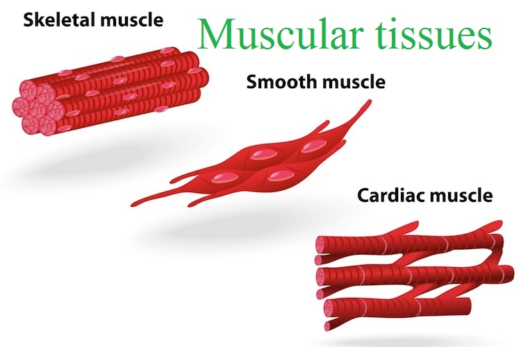
Muscular tissues can be classified into: Smooth, non-striated or involuntary muscles. Cardiac muscle or myocardium. Skeletal, striated or voluntary muscles. Skeletal muscles: Skeletal muscle comes in different shapes and sizes and allows movement of the body and body parts. These muscles have the longest fibers. Skeletal muscles have striations and can be controlled voluntarily. Skeletal muscles are able to contract very rapidly but they tire easily; they must also rest after relatively short periods of activity otherwise physical damage and muscle fatigue will occur. Skeletal muscles can exert tremendous power and are remarkably adaptable. Skeletal muscle is attached to the skeleton. The movement of these muscles also cannot be controlled by will. Smooth muscle Smooth muscle tissue is found in the walls of the hollow visceral organs such as the respiratory passages, intestinal tract and urinary bladder. This is not striated and is involuntary. Contractions of smooth muscles are slow and sustained. The contraction of most of these muscles forces fluid and other substances through the internal body channels. Smooth muscle contractions are involuntary movements triggered by impulses that travel through the autonomic nervous system to the smooth muscle tissue. The arrangement of cells within smooth muscle tissue allows for contraction and relaxation with great elasticity. The smooth muscle in the walls of organs like the urinary bladder and the uterus allow those organs to expand and relax as needed. The smooth muscle of the alimentary canal (the digestive tract) facilitates the peristaltic waves that move swallowed food and nutrients. In the eye, smooth muscle changes the shape of the lens to bring objects into focus. Artery walls include smooth muscle that relaxes and contracts to move blood through the body. Cardiac muscle: Cardiac muscle makes up most of the heart walls. The muscles are striated and are involuntary – we have almost no conscious control over how our heart beats. The heart wall is composed of three layers. The middle layer, the myocardium, is responsible for the heart’s pumping action. Cardiac muscle found only in the myocardium, contracts in response to signals from the cardiac conduction system to make the heartbeat. Cardiac muscle is made from cells called cardiocytes. Cardiac muscle cells usually contract at a steady pace set by the heart’s pacemaker cells, though nerves can influence them to speed up or slow down. The cardiac muscle tissue forms many branches, which then recombine to form a continuous sheet. This enables the tissue to contract as a unit and improves its efficiency. Cardiac muscle never rests – to do so would be fatal. Contractions of cardiac muscle move blood through the heart and around the body.
Cardiovascular system
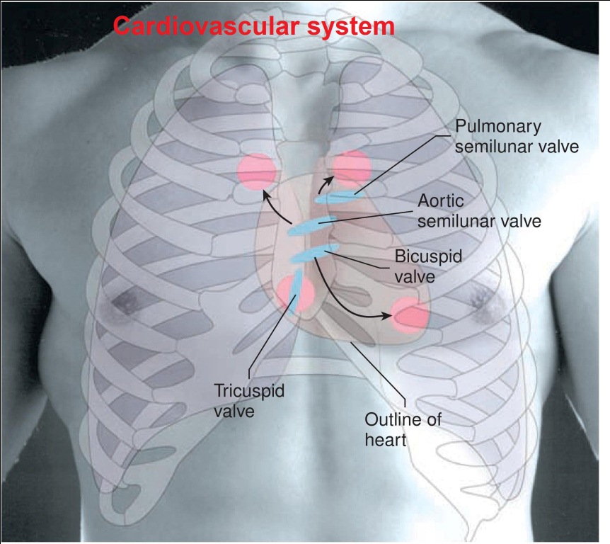
Cardiovascular system Humans have a closed circulatory system. You may remember that in a closed system blood is contained within vessels and that the main components of a closed circulatory system are the heart, blood vessels, and blood. Heart The heart is a conical, hollow, musculotendinous organ. It lies in the thorax between the lungs and behind the sternum. It’s about 10cm long and weighs about 300 gram. The base of the heart is above and the apex is below. The heart lies in the thorax between the lungs and behind the sternum. Two-thirds of the heart is on the left side. It lies obliquely. It’s directed more towards the left side than on the right side. The apex of the heart lies at the level of 5th intercostals space, 9cm to the left of midline. The base extends to the level of the second rib. The 3 Layers of Heart: The heart is surrounded by an outer covering called pericardium. It contains two layers called visceral pericardium and parietal pericardium. Pericardial fluid is present between these two layers. The middle layer is made of heart muscle fibers. It’s called as myocardium The inner lining is called as endocardium. Chambers of the heart: The Human heart has four chambers: two atria and two ventricles. The 2 chambers on the right side are known as Right Atrium, Right Ventricle. The 2 chambers on the left side are called Left Atrium, Left Ventricle. The 4 chambers are divided by 2 septa, the interatrial septum, and Inter-Ventricular septum. Valves of the heart: The heart has 4 valves: they are as follows The mitral valve and tricuspid valve, which controls blood flow from the atria to the ventricles. The aortic valve and pulmonary valve, which control blood flow out of the ventricles. Some tendinous cords arise from the lower border of these valves. They’re called chordate tendinae. The chordate tendinae in turn attached to papillary muscle which arises from ventricular walls. Blood vessels attached to heart: the right atrium receives superior vena cava vein and inferior vena cava vein. They carry venous blood to the heart. From the right ventricle, arise the pulmonary artery. It carries venous blood to lungs for oxygenation. The left atrium receives 4 pulmonary veins. They carry oxygenated blood to the heart. From the left ventricle. Arise the aorta. It delivers pure-blood to all parts of the body. Blood supply to the heart: The heart receives its blood supply through right and left coronary arteries. They’re the first branches of the aorta. Venous blood heart is collected by the coronary sinus. It opens directly into the right atrium. Nerve supply to the heart: The heart is supplied by sympathetic and vagus nerves. Branches from these nerves pass through the sino auricular node.
BONE DEVELOPMENT
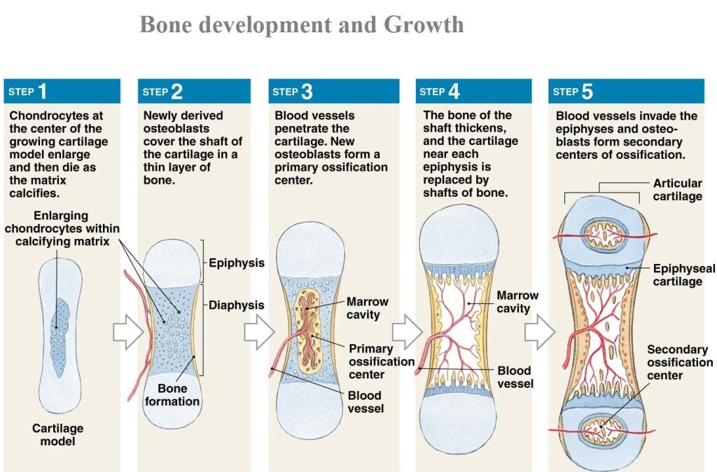
BONE DEVELOPMENT: There are two processes that form our bones before we are born: intramembranous ossification and endochondral ossification. Bones of the skeleton are developed in 2 ways they are as follows: Intramembranous ossification: Intramembranous ossification is primarily responsible for forming the bones in our skull, and bones are formed from a specific type of connective tissue, called mesenchymal connective tissue. The replacement of sheet-like connective tissue membranes with bony tissue. Bones formed in this manner are called intramembranous bones. They include certain flat bones of the skull and some of the irregular bones. The future bones are first formed as connective tissue membranes. Osteoblasts migrate to the membranes and deposit bony matrix around themselves. When the osteoblasts are surrounded by matrix they are called osteocytes. Endochondral ossification is the process by which the embryonic cartilaginous model of most bones contributes to the longitudinal growth and is gradually replaced by bone. Endochondral ossification involves the replacement of hyaline cartilage with bony tissue. Bone Growth: Bone growth happens at the ends of the bones at the growth plate. As the bones grow, cells in the growth plate first produce more cartilage to extend the bone and then osteoblasts come in and convert the cartilage to bone in the same way that they do when bones are first formed before birth. Bones can continue to grow until about the age 16 to 20 when the growth plate turns completely to the bone and no new cartilage can be added at the ends of the bones.
Abnormal Breathing and Breathing Disorders:
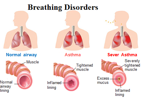
Abnormal Breathing and Breathing Disorders Apnea:Absence of breathing. Orthopnea:Only able to breathe comfortably in upright, unable to breath laying down Dyspnea: Subjective sensation related by patient as to breathing difficulty Paroxysmal nocturnal dyspnea – attacks of severe shortness of breath that wake a person from sleep, such that they have to sit up to catch their breath – common in patients with congestive heart failure. Hyperventilation: Hyperventilation occurs when the rate and quantity of alveolar ventilation of carbon dioxide exceed the body’s production of carbon dioxide. Hypoventilation: Hypoventilation occurs when ventilation is inadequate to perform needed gas exchange. Hypoventilation is considered a precursor to hypoxia and its lethality is attributed to hypoxia with carbon dioxide toxicity. Cheyne-Stokes respirations (CSR): Gradual increase in volume and frequency, followed by a gradual decrease in volume and frequency, with apnea periods of 10 – 30 seconds between cycle. Described as a crescendo – decrescendo pattern. Characterized by cyclic waxing and waning ventilation with apnea gradually giving way to hyperpneic breathing.
Autonomic Nervous System
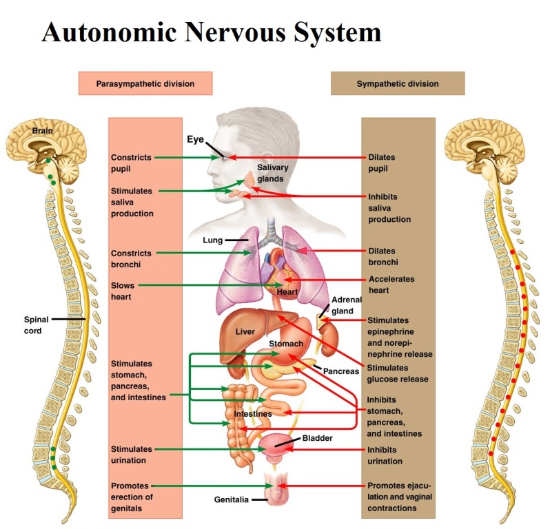
Autonomic Nervous System The autonomic system is the part of the peripheral nervous system responsible for regulating involuntary body functions, such as blood flow, heartbeat, digestion, and breathing. In other words, it is the autonomic system that controls aspects of the body that are usually not under voluntary control. This system allows these functions to take place without needing to consciously think about them happening. Autonomic Nervous System is further divided into two branches: The sympathetic system regulates the flight-or-fight responses. This system prepares the body to expend energy and deal with potential threats in the environment. When the action is needed, the sympathetic system will trigger a response by speeding up the heart rate, increasing breathing rate, increasing blood flow to muscles, activating sweat secretion, and dilating the pupils. This allows the body to respond quickly in situations that require immediate action. In some cases, we might stay and fight the threat, while in other cases we may instead flee from the danger. The parasympathetic system helps maintain normal body functions and conserve physical resources. Once a threat has passed, this system will slow the heart rate, slow breathing, reduce blood flow, to muscles and constrict the pupils. This allows us to return our bodies to a normal resting state.
Muscle Physiology
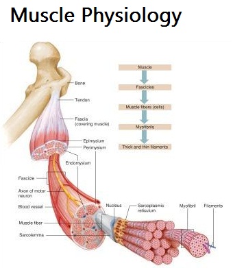
Muscle Physiology Muscle cells are specialized to contract. The cells are similar in structure to the other cells in the body but are elongated and, for this reason, are called muscle fibers. They consist mostly of water (about 75%), proteins and inorganic salts. There are around 700 different muscles in the human body and more than 250 million muscle fibers. The body contains three distinct types of muscle (skeletal, cardiac and smooth), which will be outlined in this article. The following three articles in the series will address the structure and function of skeletal muscle, the largest muscle mass in the body. Muscles are excitable or irritable. This means that they are capable of receiving and responding to a stimulus. The stimulus is usually a chemical – a neurotransmitter released by a nerve cell, a hormone or a local change in acidity (pH). In response, muscles generate an electrical impulse that causes the muscle cells to contract: – Muscles can contract or shorten when they are stimulated. No other type of body tissue can do this. – Muscles are extensible – that is, they can stretch or extend beyond their resting length. – Muscle fibers are elastic and can return to their resting length after being stretched.
Blood pressure (BP)
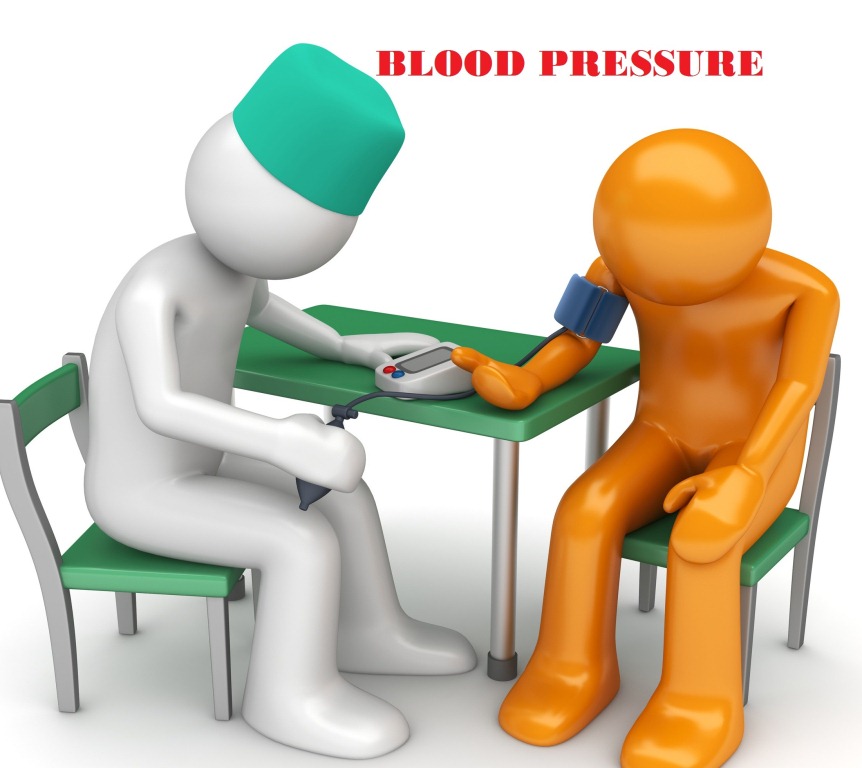
Blood pressure (BP) It’s the lateral pressure excreted by blood on blood vessels. It’s normally expressed as arterial pressure. It has 2 phases: Systolic blood pressure: it’s the maximum pressure occurs during the systole of the heart range from 100 to 120 mm Diastolic blood pressure: it’s the minimum pressure occurs during the diastole of the heart range between 60 to 80 mm Hg. Pulse pressure is the difference between systolic and diastolic blood pressure and it’s nearly 40 mm Hg.
Vajrasana – Thunderbolt Posture
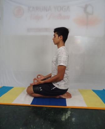
Vajrasana – Thunderbolt Posture (Vajra – thunder bolt or diamond) Practice: Come to (Dandasana) staff posture, by palms beside hip. Slide towards left, bend at right leg knee, and rest it under right hip. Same, bend left leg knee, place it under left hip. Keep spine, neck, head straight. Heel contacting with hip, toes pointing outward, inner calf muscles touching quadriceps muscle, knees together. At fingers adopt Jnana Mudra by touching tip of index, thumb finger slightly and rest it on respective thigh. Stay for 10 to 40 seconds, breathing completely by utilizing abdomen and chest area. Return back, sliding towards right side, with palm support unfold both legs and relax. Benefits: This is the only one asana, immediately after food we can perform to aid digestion by stimulating Vajra Nadi. Give suppleness to ankles, reduce thigh, hip extra fat. Make spine straight and strong naturally. It induces meditative state, so good for meditation.
Utkatasana – Mighty Posture
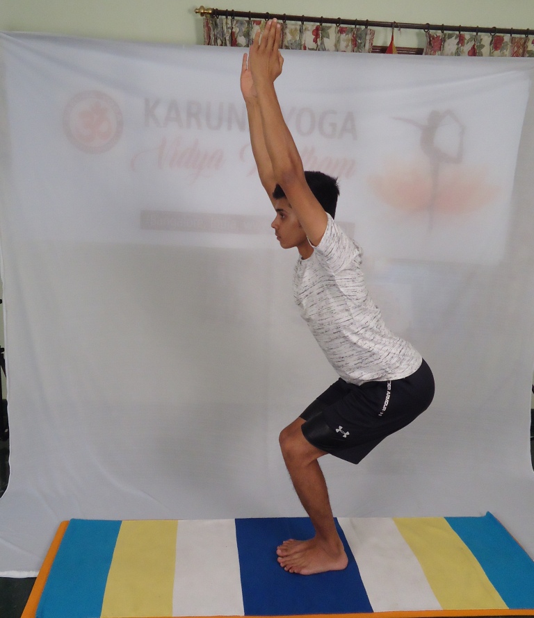
Utkatasana – Mighty Posture (utkata = mighty or powerful) Practice: Come to Tadasana, by keeping legs together and palms beside your thighs. As you inhale raise your arms till your biceps touch ears, palms together finger pointing towards sky, lock your elbow and keep straight, tuck your elbows, open your chest and expand , rib cage lifted up, abdomen in , keep your entire spine extended and lengthen up. Exhale and bend your knees , squatting down until your thighs are parallel to the floor, do not fall forward, move backward let your weight distribute over at lower back , hip joints and thigh muscles. Breathing normally at your nose, slow, even, and long breathing. Stay in this posture for 10 to 30 seconds or till you fill comfortable, do not exert or overstrain your body, always listen to your body. Along exhalation bring arms beside your thighs, and inhale straightens your knees. Again come back to Tadasana and relax. Benefits: Strengthens shoulders and open the chest. Strengthens and tones the ankles, thighs, calves, and spine Activate and optimize the efficiency of abdominal organs, diaphragm, and heart. Reduces flat feet.