Respiration Mechanism
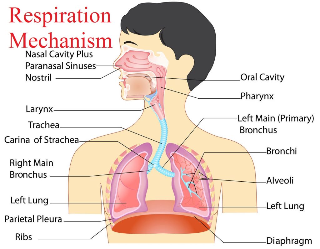
Respiration mechanism Normal breathing involves several different mechanisms. Shallow breathing is accomplished by the contraction of the diaphragm and the external intercostal muscles for inhalation. During exhalation, the muscles relaxing and the elasticity of the lungs returning to their resting volume expels air out of the lungs. Deep Breathing is accomplished by an inferior movement of the diaphragm towards the abdomen. The external intercostal muscles along with the sternocleidomastoid and scalene muscles in the neck expand the space between the ribs, increasing the volume of the chest. During deep exhalation, the internal intercostal muscles and abdominal muscles contract to decrease the volume of the thoracic cavity, forcing air out of the lungs. Breathing is controlled by the brain and may be controlled both consciously and unconsciously. Unconscious control of breathing is maintained by the respiratory center of the brainstem, which monitors the concentration of gases in the blood and adjusts the rate and depth of breathing as needed. During Yoga practice, the respiratory center automatically increases the breathing rate to provide constant levels of oxygen to the blood. During rest, the respiratory center reduces the breathing rate to prevent hyperventilation and maintain healthy oxygen and carbon dioxide levels in the blood. Conscious control of breathing is maintained by the cerebral cortex of the brain. The cerebral cortex may override the respiratory center during the practice of Pranayama. Unconscious control of breathing resumes as soon as conscious control of breathing ends, preventing the body from suffocating from lack of breathing.
Peripheral Nervous System

Peripheral Nervous System: The Peripheral nervous system (PNS) is the division of the nervous system containing all the nerves that lie outside of the central nervous system (CNS). The primary role of the PNS is to connect the CNS to the organs, limbs, and skin. These nerves extend from the central nervous system to the outermost areas of the body. The peripheral system allows the brain and spinal cord to receive and send information to other areas of the body, which allows us to react to stimuli in our environment. The nerves that make up the peripheral nervous system are actually the axons or bundles of axons from neuron cells. In some cases, these nerves are very small but some nerve bundles are so large that they can be easily seen by the human eye.
Skeletal Muscle Structure
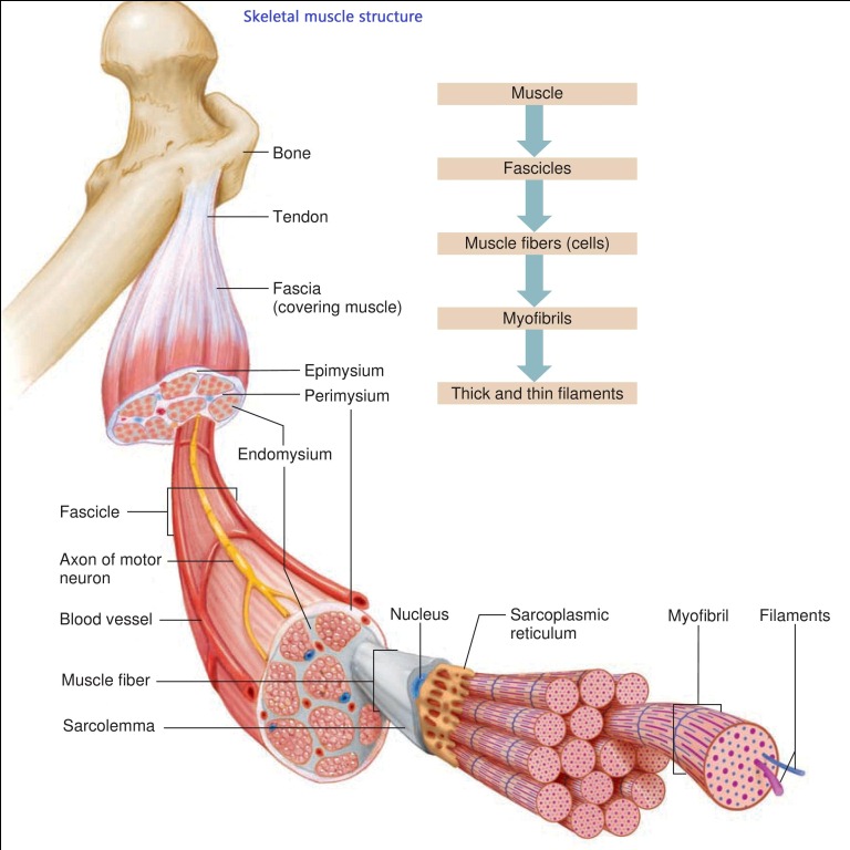
Skeletal Muscle Structure Skeletal muscles are composed of a large number of muscle fibers. Each muscle fiber has no one or more nuclei which lie in the periphery. The cytoplasm of the muscle cell is as sarcoplasm. Skeletal muscles are composed primarily of contractile material. Skeletal muscle is a composite tissue of connective tissue, blood vessels, and nerves as well as contractile material, these “minor” tissues may strongly influence muscle function. Myofibrils are very small parallel filaments which lie in the cytoplasm. The membrane of the muscle fiber is called as sarcolemma. Each muscle fiber is embedded in a connective tissue called endomysium. The fibers within a muscle are arranged in bundles. Each bundle is enclosed in a sheath called perimysium.
Conducting system of the heart

Conducting system of the heart Conducting system of the heart: the impulse for cardiac contraction is transmitted through the conduction system of the heart. This system is made of : Sino atrial node (SA-node) Atrioventricular node (AV-node) Bundle of his Purkinje fibers. Heart sounds: Totally 4 sounds are produced by the heart. The first sound as LUB and the second sound as DUB can be heard with a stethoscope. The third and fourth sounds cannot be heard. The “ lub” is the first heart sound, commonly termed S1, and is caused by turbulence caused by the closure of mitral and tricuspid valves at the start of systole. The second sound, “dub” or S2, is caused by the closure of aortic and pulmonic valves, marking the end of systole. Heart Murmurs. The most common abnormal heart sound is a heart murmur. A murmur is a blowing, whooshing, or rasping sound that occurs during your heartbeat. Pulse: its throbbing sensation felt over the walls of arteries. It’s defined as the pressure difference transmitted in the form of a wave over the arterial walls. Heart Rate: the normal resting heart rate for adults ranges from 60 to 100 beats a minute. The wrist is the common site where the pulse is usually felt. At this site, the radial artery is very superficial. This pulse is called as a radial pulse. Other arteries where a pulse can be felt are a carotid artery. Facial artery and temporal artery. In case of fever, the rate of pulse increases at the rate of 10 per every rise of 1F.Generally, veins don’t exhibit pulsation. The only vein which exhibits pulsation is jugular vein which is nearer to the heart.
Classification of Human Bones
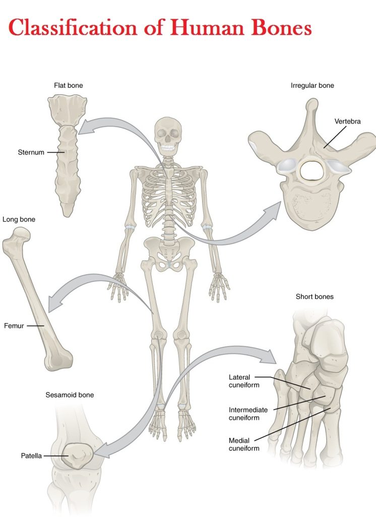
Classification of Bones according to shape Long bones: Long bones are characterized by a long tubular shaft and an articular surface at each end of the bone where ligaments and tendons attach. They’re found in the limbs to facilitate movement and support the weight of the body. These bones include the major bones of the arms and legs such as the humerus and femur, tibia and fibula, and the radius and ulna. Short bones: Short bones are roughly cube shaped and are as long as they are wide. It has no shaft but it contains a spongy substance covered by a shell of the compact. Helps to provide stability and movement within the ankle and wrist joints. They provide little to no movement. Examples of this type of bone include the carpals and metacarpals in the wrists and ankles. Flat bones: The primary purpose of this type of bone is to protect internal organs such as the brain, heart, and lungs. It also provides a large surface area for muscles to attach to. Examples of this type of bone include the cranium (skull), the thoracic cage (sternum and ribs) and the ilium (pelvis). These contain two layers of compact bone with a spongy substance in between. Irregular bones: This type doesn’t fall in any category e.g. vertebrae and the bones of the face. These bones vary in size and structure with the shape usually being very complex. Irregular bones serve different functions depending on location. Sesamoid bones: they are small bones which develop in the tendons of muscles e.g. patella of the knee joint. The function of this bone is to protect tendons and diminish friction and wear on joint surfaces.
Pulmonary Ventilation
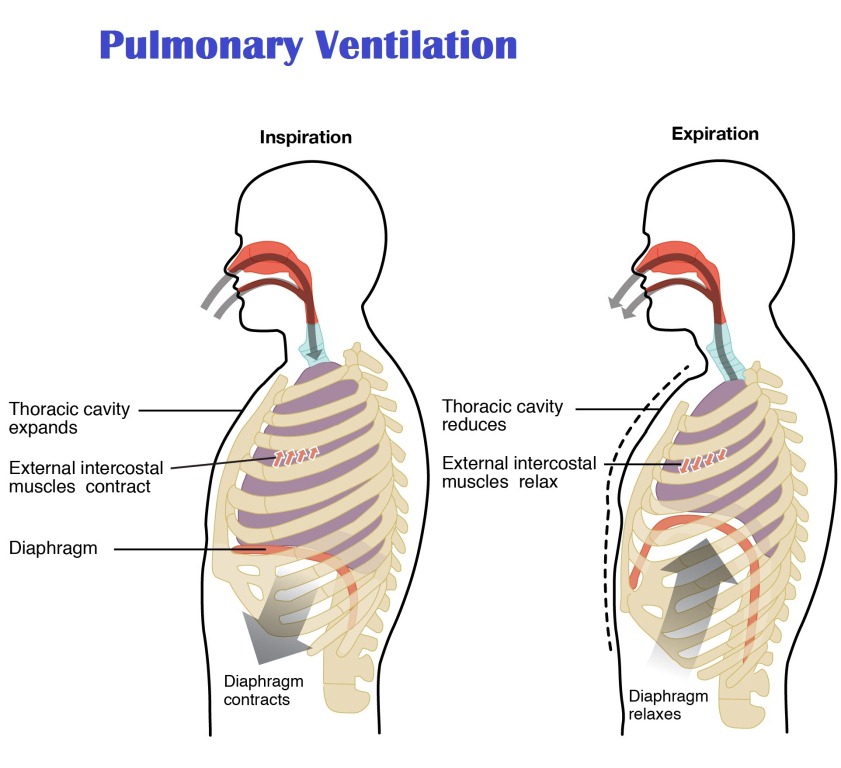
Pulmonary Ventilation Smooth muscle cells in the walls of the bronchioles adjust their diameter and help to control the flow of air into the alveoli of the lungs. The muscles of the thoracic cavity, including the diaphragm and intercostal muscles, change the volume of the thoracic cavity to force air into and out of the lungs. The diaphragm contracts and pushes itself into the abdominal cavity, increasing the volume of the thoracic cavity and expanding the lungs to draw in fresh atmospheric air. When it relaxes, the diaphragm returns to its original position and forces air out of the lungs during exhalation. The intercostal muscles also aid in respiration by lifting the ribs during deep inhalation to increase the size of the thoracic cavity and by pushing the ribs together during deep exhalation to decrease the thoracic cavity.
Nervous System
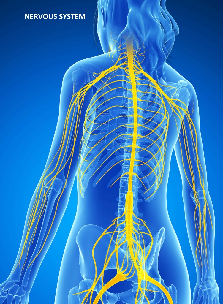
Nervous System The nervous system has been divided into two components: The central nervous system which is composed of the brain and the spinal cord, and the peripheral nervous system, which is composed of ganglia and peripheral nerves that lie outside the brain and spinal cord. The peripheral nervous system has been, in turn, divided into two subsystems: Somatic and autonomic. Somatic Nervous system (SNS): The somatic nervous system is responsible for the movement of voluntary muscles and the process known as a reflex arc. This system carries nerve impulses back and forth between the central nervous system and the skeletal muscle’s, skin and the sense organs. Autonomic Nervous system (ANS): The Autonomic Nervous system controls and regulates the internal organs without any conscious recognition or effort by the organism.
CHARACTERISTICS OF SKELETAL MUSCLES
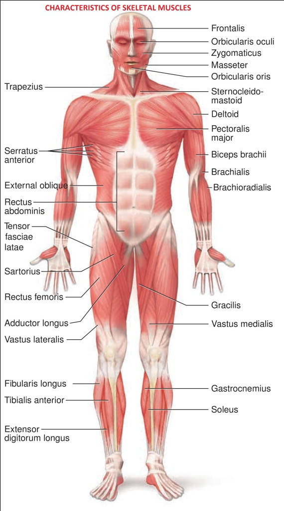
CHARACTERISTICS OF SKELETAL MUSCLES The four major functional characteristics of skeletal muscle are: Contractility – The ability to shorten which causes movement of the structures to which the muscles are attached. Excitability – The ability to respond or contract in response to chemical and/or electrical signals. Extensibility – The capacity to stretch to the normal resting length after contracting Elasticity – The ability to return to the original resting length after a muscle has been stretched. Functions of Muscles: Movement: Skeletal muscles move bones by pulling on them and work together to produce smooth movements of our limbs. Mobility is your muscular system’s simplest and most crucial function. Your skeletal muscles are largely responsible for the movements and motions you make. Skeletal muscles are attached to your bones. Maintaining posture, muscle tone, and Stabilizing joints: Skeletal muscles also control posture, from your head down to your toes. Flexibility and strength are keys to maintaining proper posture. Stiff neck muscles, weak back muscles, and tight hip muscles, among other ailments, can throw off your alignment. Producing Heat & Temperature Regulation: most of the heat required for this is produced by muscles, which generate heat as they contract. Skeletal muscle accounts for at least 40% of body mass and is the muscle type responsible for generating most body heat. Circulation: The involuntary cardiac and smooth muscles help your heart beat and blood flow through your body. The cardiac muscle, known as the myocardium, is found in the walls of the heart. The cardiac muscle is controlled by the autonomic nervous system, which is responsible for most bodily functions.
Cardiac Output

Cardiac Output The cardiac output is simply the amount of blood pumped by the heart per minute. Necessarily, the cardiac output is the product of the heart rate, which is the number of beats per minute, and the stroke volume, which is amount pumped per beat. Stroke volume is the amount of blood ejected per beat of the heart. It’s about 70ml. so every mixture about 5040 ml nearly 5 liters of blood is pumped by the heart. Cardiac output depends on the following factors:
Bone structure and formation
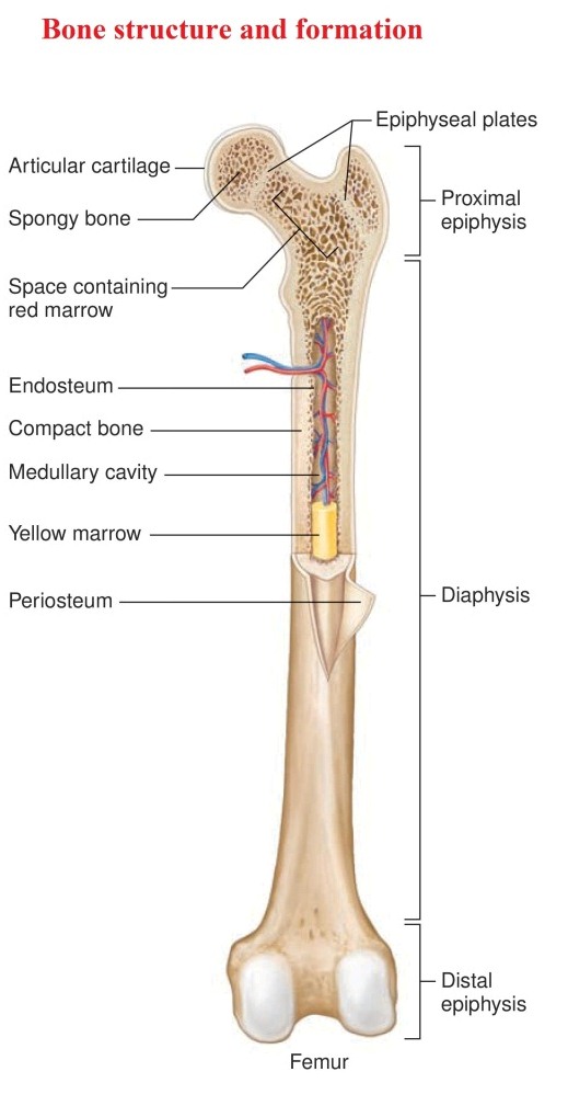
Bone structure and formation: Bone is the hardest of the connective tissues. Compact bone (Cortical Bone) and Spongy bone (Spongy Bone) are the two types of osseous tissue or bone tissue that make up bones. The bones are mostly composed of an outer layer of compact bone and an inner layer of spongy bone. Ossification is the process of bone formation. Parts of the skeleton form during the first few weeks after conception. By the end of the eighth week after conception, the skeletal pattern is formed in cartilage and connective tissue membranes and ossification begins. Compact bone (Cortical Bone): Due to the strength of compact bone, its main functions is to support the entire body. Compact pact bone also stores calcium. While calcium is being stored in the bone it is also the principle molecule that hardens compact bone. Cancellous bone (Spongy Bone): It looks spongy and found in the ends of the long bones, short bones and in between two layers of compact tissue of flat bones. Due to the sponge-like or porous nature of spongy or cancellous bone, spongy bone has a greater surface area compared to compact bone. This allows the bone marrow to develop in the region of the spongy bone. The branching nature of spongy bone also makes it a prime target in diseases such as osteoporosis.