Why yoga student should know about “human anatomy and physiology”
Understanding yoga anatomy is crucial for all yoga students because it will increase the effectiveness of the asanas and knowledge to protect from creating injuries and also be better prepared to deal with any existing injuries safely and effectively.
Importance of studying anatomy in hatha yoga application
Importance of studying anatomy in hatha yoga application Anatomy: Anatomy is the study of the structure of an organism. This involves the understanding of appearance, structure, location, organization of the various parts of the organism. Importance of studying anatomy in yoga: In Ashtanga Yoga, the practice of asanas and pranayama engages the entire body and physiological processes. They understand the right way to perform an asana the structure is needed to be clear which requires the understanding of bone and muscle alignment. An understanding of anatomy also gives the individual the efficiency to analyze the practice to create effective movement patterns. This effective practice will further aid and prevent the body from possible injuries. Furthermore, knowing one’s body helps in understanding one’s body better, bringing the focus of one’s awareness to the area of the body which requires more attention. In summary, it helps in being more tuned to one’s own body. In pranayama practice, anatomy helps in understanding the movement of prana (breath) within the body. Understanding where the breath is traveling during different pranayama practices. For example, the movement of the abdomen during abdomen breathing and movement of ribs/ chest during thoracic breathing. It facilitates the focus needed during pranayama. Physiology: It is the study of the process of living organisms. Study of how the organism functions. Cell: It is the smallest unit responsible for all life’s processes. Systems in the Human Body: Locomotor System Blood Vascular System Digestive System Respiratory System Ductless System Urogenital System Nervous System Special Sense Organ System Excretory System Locomotor System: It consists of parts of the body responsible for movement including the skeletal system, bones, cartilages, ligaments, muscular system, etc. Blood Vascular System: It is the lymphatic or circulatory system, it consists of the heart, arteries, and veins. It is responsible for the circulation of nutrients, blood, oxygen, etc to various parts of the body. It is also responsible for removing waste from the blood and the lymph, it is responsible for fighting diseases and hence is important for immunity. Respiratory System: It is a network of organs and tissues that function together to exchange oxygen and carbon dioxide. Releasing carbon dioxide and In taking oxygen. It consists of the nose, nasal cavity, pharynx, larynx, trachea, bronchi, bronchioles, and lungs. Nervous System: It is a complex network of the brain, spinal cord, nerves, neurons, and other non- neuron cells and sensory receptors that are responsible for several functions including perception, sensation, movement, thinking, formation of memories, emotions, etc. Courtesy To: Shrutika Mishra, RYT – 200 Hour
YOGA AND ENDOCRINE GLANDS

Heart, Arteries & Veins Forms the Circulatory System
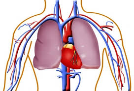
The Heart, Arteries & Veins Forms the Circulatory System: The heart pumps blood into arteries. The arteries divide and subdivide and finally end in capillaries. The capillaries later unite to form veins. The veins return blood to the heart. The arteries carry pure blood away from the heart, Veins carry De-oxygenated blood to the heart and the capillaries are minute channels receives blood from smaller arteries (arterioles) and deliver into smaller veins (venules). Blood circulation: depending on the course of blood. Circulation can be classified into: Systemic circulation. Pulmonary circulation Coronary circulation Portal circulation. Systemic circulation: it’s the blood circulation that carries oxygenated blood away from the heart, to the body, and returns deoxygenated blood back to the heart except for lungs. This circulation starts Oxygenated blood enters the left atrium from the pulmonary veins. The blood is then pumped through the mitral valve into the left ventricle. From the left ventricle, blood is pumped through the aortic valve and into the aorta, the body’s largest artery. The aorta arches and branches into major arteries and then it breaks up into smaller arteries and finally ends in capillaries. The capillaries unite to form venules which join up ultimately to form 2 large venous trunks namely superior vena cava and inferior vena cava. These 2 venous trunks open in the right atrium of the heart. Gas and nutrient exchange with the tissues occurs within the capillaries that run through the tissues. Metabolic waste and carbon dioxide diffuse out of the cell into the blood, while oxygen and glucose in the blood diffuse out of the blood and into the cell. The arterial component of systemic circulation the highest blood pressures in the body. The venous component of systemic circulation has considerably lower blood pressure in comparison, due to their distance from the heart, but contain semi-lunar valves to compensate. Systemic circulation as a whole is a higher pressure system than pulmonary circulation. Pulmonary circulation: it involves the purification of blood in lungs. It’s the circulation of the blood from the heart to the lungs for oxygenation, then back to the heart again. The Oxygen-depleted blood from the body leaves the systemic circulation when it enters the right atrium, The blood is then pumped through the tricuspid valve into the right ventricle. From the right ventricle, blood is pumped through the pulmonary valve and into the pulmonary artery. The pulmonary artery splits into the right and left pulmonary arteries and travel to each lung. The oxygenated blood then leaves the lungs through pulmonary veins, which returns it to the left atrium, completing the pulmonary circulation. Coronary Circulation: Coronary circulation involves blood supply to the heart itself, it is the circulation of blood in the blood vessels of the heart muscle. The vessels that deliver oxygen-rich blood to the myocardium are known as coronary arteries. The right and left coronary arteries arise from ascending aorta. The vessels that remove the deoxygenated blood from the heart muscle are known as cardiac veins which collect and opens into the right atrium. Portal circulation: It’s the circulation of blood through the liver. In this circulation, the portal vein carries blood that has circulated in stomach, intestine, and pancreas to liver. The portal vein divides into capillaries. These capillaries join with the capillaries of the hepatic artery. The venous blood of liver is collected by hepatic vein which joins with inferior vena cava.
Measurement of blood pressure
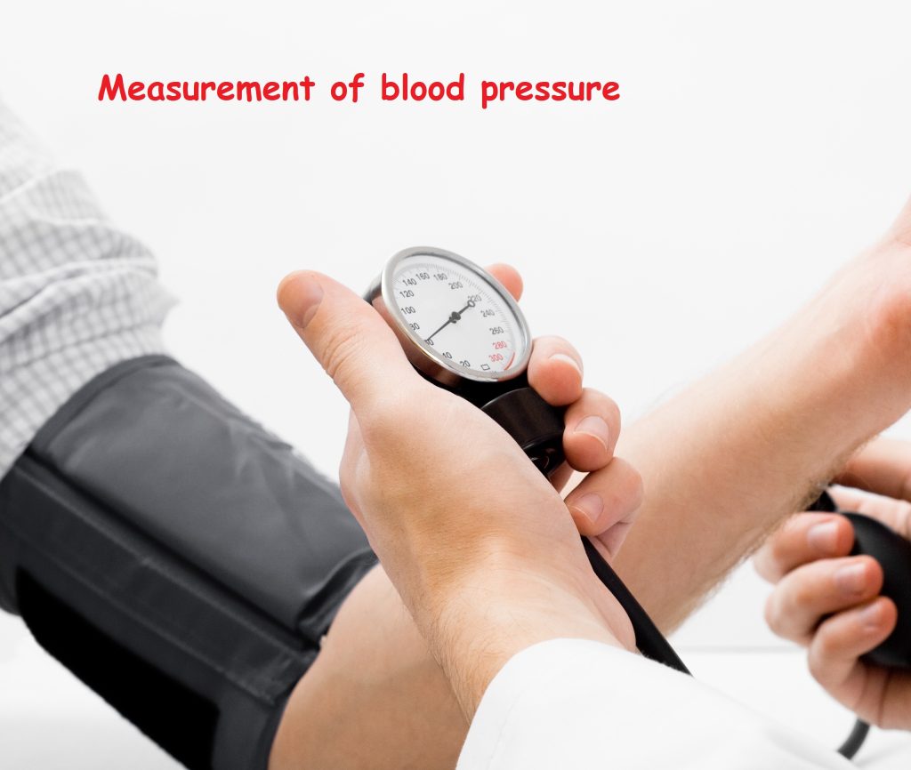
Measurement of blood pressure: Blood pressure is usually measured by an instrument called sphygmomanometer; it consists of a mercury manometer. Cuff and hand pump. The cuff is tied around the cubical fossa of the individual then the hand pump is pressed so that air is inflated in the cuff. When the cuff is fully inflated, air pressure is more than blood pressure. So blood flow in the brachial artery is completely obstructed. Now the hand pump is slowly released until the time the appearance of the first sound is heard (by means of a stethoscope put in the cubital fossa*. The manometer reading is now noted this regarding the systolic pressure. Later the hand pump is slowly released until the time that the sound becomes louder and louder. Later it stops the manometric reading is noted when the sound disappears. This reading is the diastolic blood pressure.
Main bones of the human skeleton
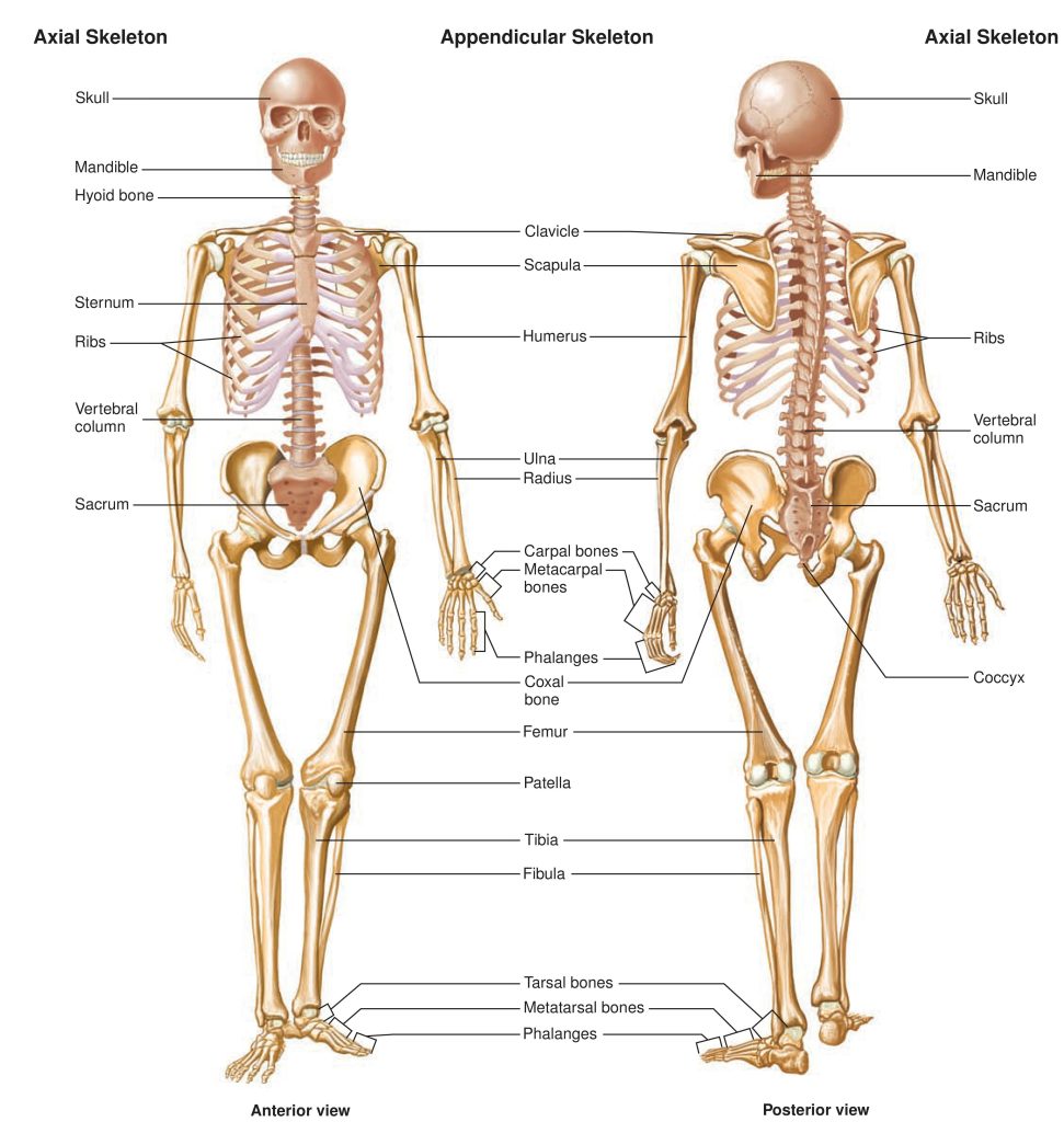
The main bones of the human skeleton are: The Skull – Cranium, Mandible, and Maxilla Shoulder girdle – clavicle and scapula Arm – humerus, radius, and ulna Hand – Carpals, Metacarpals, and Phalanges Chest – Sternum, and Ribs Spine – Cervical area (top 7 vertebrae), Thoracic (next 12), Lumbar (bottom 5 vertebrae), Sacrum (5 fused or stuck together bones) and Coccyx (the tiny bit at the bottom of the spine). Pelvic girdle – Ilium, Pubis, and Ischium. Leg – Femur, Tibia, and Fibula Ankle – Talus and calcaneus Foot – Tarsals, Metatarsals, and Phalanges.
Factors affecting blood pressure
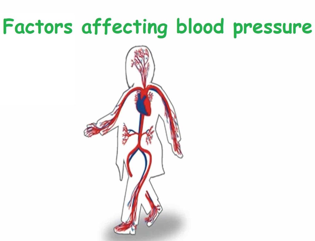
Factors affecting blood pressure: Blood volume. Cardiac output Peripheral resistance. The elasticity of blood vessels. The diameter of the lumen of blood vessels. The viscosity of blood. Blood volume: it’s the total amount of blood in circulation. A sufficient amount of blood in blood vessels is necessary to maintain normal blood pressure loss of blood as in hemorrhage produces a fall in blood pressure. Cardiac output is the quantity of blood pumped by the heart in one minute. It’s the product of stroke volume and the heart rate. An increase in stroke volume increases systolic blood pressure. An increase in cardiac output increases both systolic and diastolic blood pressure. Peripheral resistance: is the resistance offered by blood vessels for the flow of blood. Resistance is offered mainly by small blood vessels, especially arterioles. Elasticity of the arterial walls distends the aorta when the ventricle contracts. The elastic recoils when the ventricle relaxes. This recoiled pushes the blood onwards. The decrease in elasticity as in atheroma produces a rise in blood pressure. 50 The diameter of the lumen of blood vessels: can be altered, narrowing of the lumen increases the resistance to blood flow and this increases blood resistance to blood flow and this increases blood pressure. Enlargement of the lumen has the opposite effect. The viscosity of blood: it’s the blood stickiness. The viscosity of blood spends on plasma, plasma proteins and number of the red blood cells. An increase in viscosity increases blood pressure.
Disorders of Heart
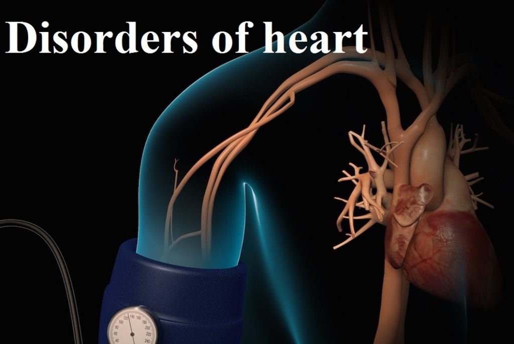
Disorders of Heart Cardiac failure: It’s a condition in which the myocardium of ventricle is unable to maintain sufficient circulation of blood to meet the needs of the body. Depending on onset it may be classified into: 1- Acute failure: when it’s sudden. 2- Chronic failure: when it’s gradual. Stenosis of valves: it’s the narrowing of the valves of the heart. In this condition, the edges of the cusps of the valves become rough. So they stick together and narrow the valvular opening. The incompetence of valves: it’s a functional defect caused by the failure of the valve to close completely. This allows blood to flow back into the ventricle when it relaxes. Angina pectoris: it’s a pain occurring due to myocardial ischemia. It occurs due to narrowing of coronary arteries. Due to this, physical effort causes severe ischemic pain. Myocardial infarction: it’s the death of an area of cardiac tissue due to lack of coronary blood supply to the segment of the myocardium. It occurs due to occlusion of the coronary artery. Cardiac arrhythmia: it’s a disorder in cardiac rate and rhythm. It occurs due to defective impulses formation and defective impulse conduction in the heart.
Electrocardiogram ECG
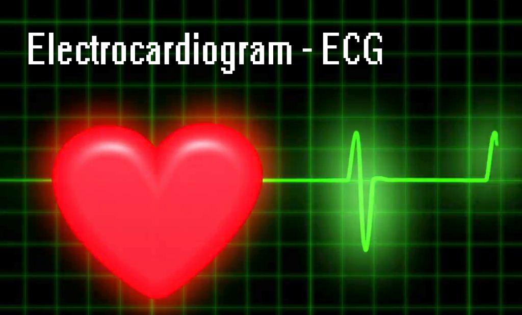
Electrocardiogram ECG: The ECG shows the heart’s electrical activity as line tracings on paper. It’s the recording of electrical activity of the heart. ECG is an instrument which is used to record the electrical current generated in the heart. By means of this instrument, the electrical current generated in the heart is conducted to remote by connecting any two parts of the body with this instrument. The connections are called as leads they are. A normal ECG contains waves, intervals, segments and one complex, as defined below. Wave: A positive or negative deflection from the baseline that indicates a specific electrical event. The waves on an ECG include the P wave, Q wave, R wave, S wave, T wave and U wave. Interval: The time between two specific ECG events. The intervals commonly measured on an ECG include the PR interval, QRS interval (also called QRS duration), QT interval and RR interval. Segment: The length between two specific points on an ECG that are supposed to be at the baseline amplitude (not negative or positive). The segments on an ECG include the PR segment, ST segment, and TP segment. Complex: The combination of multiple waves grouped together. The only main complex on an ECG is the QRS complex. The P wave indicates atrial depolarization. The QRS complex consists of a Q wave, R wave, and S wave and represents ventricular depolarization. The T wave comes after the QRS complex and indicates ventricular repolarization.
Functions of the Skeletal System
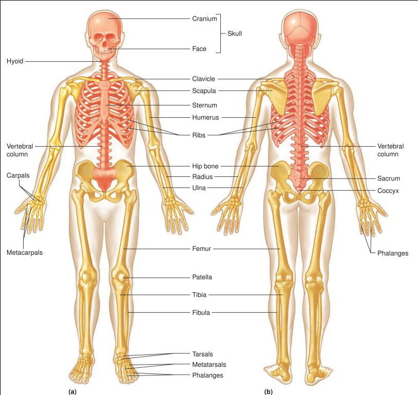
Functions of the Skeletal System? Support of the body Locomotion Provide protection for internal organs Act as a site for the production of blood cells, Calcium storage & endocrine regulation. Growth and development